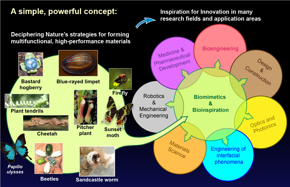
Goals and Motivation
The research in the Laboratory for Biologically Inspired Photonic Engineering is focused on the fundamental and applied aspects of conceiving and fabricating multifunctional, hierarchically-structured, bio-inspired material systems with particular focus on stimuli-responsive and dynamically tunable optical performance.
In this regard, we can benefit in several ways from highly sophisticated material solutions that have convergently evolved in various organisms. In particular, we explore design concepts found in biological photonic architectures, seek to understand the mechanisms underlying morphogenesis of biological optical materials, aim to devise viable manufacturing strategies that can benefit from insight in biological formation processes and the use of established synthetic routines alike, and ultimately strive to realize new photonic materials with tailor-made optical properties.
Projects
Our project portfolio broadly covers research in biological and bio-inspired multifunctional materials with an optical function or application potential in novel optical technology. A summary is shown in the graphic below and some project details are provided beneath:

1.
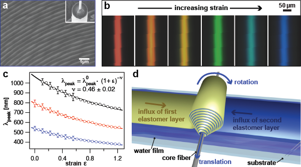
Color-tunable, stimuli-responsive photonic fibers
Project lead: Joseph Sandt (graduate student)
Contributors: Christian Argenti, Marcus Urann (undergraduate students)
This project is focused on the design, and manufacture of tunable photonic fibers. Potential applications include pressure indicators in bandages, color-dynamic textiles, infrastructural health monitoring, and applications in medical and civil engineering, where the detection of large stresses, strains, or deformations is crucial.
Related publications:
- J. Sandt et al. (in preparation).
- M. Kolle et al. Adv. Mater. 25, 2239-2245 (2013). doi-01pdf_label-01.


2.
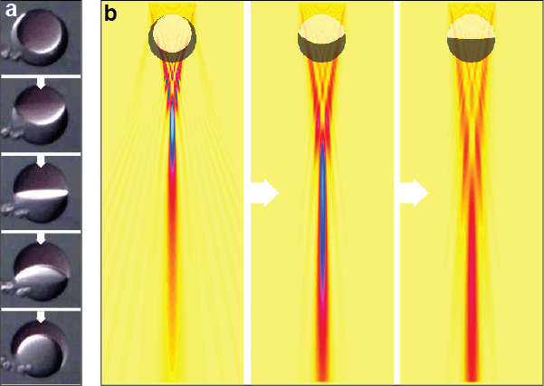
Reconfigurable, emulsion-based micro lenses
Project lead: Sara Nagelberg (graduate student)
Contributors: Lauren Zarzar (PostDoc, MIT Dept. of Chemistry), Natalie Nicolas (undergraduate student)
Tunable micro-optical elements are formed from complex emulsions. The resulting micro lenses can be controllably reconfigured using a variety of stimuli. Potential applications include stereoscopic imaging devices in microfluidics, integral imaging displays, autonomous light coupling enhancers for photovoltaics and self-adjusting fiber-couplers.
Related publications:
- J. Sandt et al. (in preparation).S. N. Nagelberg et al. (in preparation).
- L. Zarzar et al. Nature 518, 520-524 (2015).
3.
Pixelated, photonic surfaces
Project leads: Cécile Chazot, Chris Wing (graduate students)
Contributors: Maik Scherer, Alvin Tan
Hierarchical, length scale bridging arrangements of different optical features by using suitable combinations of self-assembly, template-replication and micro-fabrication, enables us to create surfaces with tens of thousands of identical optical components in one square centimeter. The visual appearance of theses surfaces changes strongly depending on the local surface orientation, which makes them suitable for localization of deformations with high spatial resolution. The incorporation of active elements such as laser dyes, and quantum dots enables us to create micro-scale emitters.
Related publications:
- S. Cormier et al., (in preparation).
- G. England et al. Proc. Natl. Acad. Sci. 111, 15630-15634 (2014).
- M. Kolle et al., Nature Nanotech. 5, 511-515 (2010).
4.
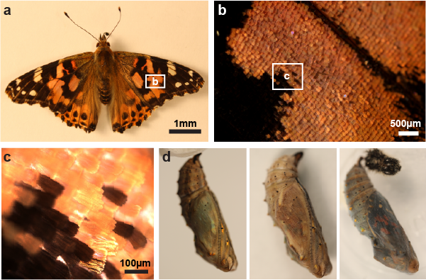
Morphogenesis and engineering of biological photonic materials
Project lead: Anthony McDougal (graduate student)
Contributors: Zahid Yaqoob (Research Scientist, MIT Laser Biomedical Research Center), William Liberti (Boston University)
Many organisms have mastered the controlled growth of hierarchical, multifunctional nano- and microstructures that show strong, optimized interactions with light. To do this, living systems employ unique physical phenomena and control mechanisms to precisely produce these functional material morphologies. We are studying the wings of the Painted Lady butterfly during metamorphosis with the aim of tracking scale development with high temporal and spatial resolution during chrysalis metamorphosis, in order to elucidate the underlying physical phenomena and control mechanisms. Our goal is to reveal key factors that we can adjust to modify structure formation in the organisms in-vivo, or in isolated cultures of scale-forming cells in-vitro.
Related publications:
- A. Dinwiddie et al., Dev. Biol. 392, 404-418 (2014).
- Y. Ohno et al., PLoS One 10, e0128332 (2015).
5.
Biomineralization of functional marine materials
Project lead: Ling Li (PostDoc, Harvard), Mathias Kolle
Contributors: Natalie Nicolas (undergraduate student)
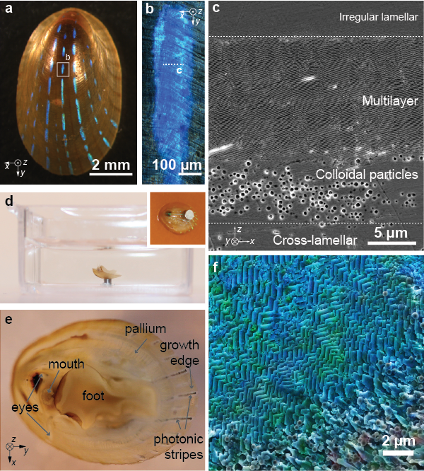
This proposed effort is aimed at revealing the mechanisms that enable the blue-rayed limpet Patella pellucida to form a translucent protective shell with an integrated photonic structure. We will use time-resolved micro-scale imaging and sensing approaches to study the formation mechanisms employed by the limpet to control the growth of its hierarchical functional shell morphology and quantify the impact that environmental parameters have on the underlying biomineralization processes. This will provide insight into low-temperature strategies for synthesizing biological and bio-inspired multifunctional materials and allow for the quantification of the influence of anthropogenic ocean acidification on the abilities of marine organisms to form shells.
Related publications:
- L. Li et. Nature Commun. 6, 6322 (2015).


